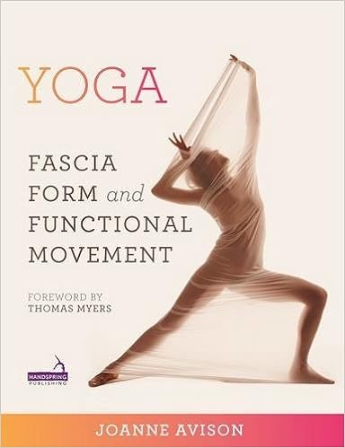
Yoga: Fascia, Anatomy and Movement
Language: English
Pages: 376
ISBN: 1909141011
Format: PDF / Kindle (mobi) / ePub
The presentation of fascial anatomy in this book provides a new context for applying knowledge of the anatomical body in a practical and relevant way to movement. Applying fascial anatomy to yoga, this book offers a way to the yoga teacher of experiencing and seeing in three dimensions - the way we really move. This enables the yoga teacher to work more creatively in the real life class.
The Yoga of Sound: Tapping the Hidden Power of Music and Chant
Yogalosophy: 28 Days to the Ultimate Mind-Body Makeover
Yoga Heals Your Back: 10-Minute Routines that End Back and Neck Pain
Weight-Resistance Yoga: Practicing Embodied Spirituality
Light on the Yoga Sutras of Patanjali
business with the branch offices of the brain, and under the general corporation law, the same as the brain itself, and why not treat it with the same degree of respect?”2 Imaginary Shapes Chapter 7 describes the standard classical biomechanical task of working from an imaginary axis, viewed as a straight pole or vertical line down the middle of the body that acts as a reference point for the sagittal, coronal and transverse planes. What happens if instead of imagining a straight pole we
which one could also state that muscle tissue is a dynamic specialisation (Ch. 5): “a ‘synovial joint’ is a contradiction in terms. It is not a joint. It is a disjoint. Here connective tissue (cartilage) enables space and therefore motion” (Jaap van der Wal).9 He went on to point out that the nature of the tissue itself made it far more than a connector. It formed “a proprioceptive substrate”, a kind of “intelligent lining” around the joint that can feel itself. It knows where it is in space via
the forming potentials in place as three layers (and three aspects on either side of centre in the middle layer), the embryo creates its length (from head to tail), its depth (from back to front) and its roundness of form by buckling (folding), taking the amniotic sac with it whilst enfolding the yolk sac. The midline of the developing notochord, neural tube and somites stiffen the back orientation (dorsal axis) supporting the embryo. The head end, the tail end and the lateral margins all grow
just being guided by bigger and better central computers (i.e. a bigger brain), they have been given many more sensory processors in the joints and fabric of the actual model. “BigDog” (Boston Dynamics) is an example of this thinking: Different branches of scientific research (neural science, developmental biology, psychology, embryology, fascia research and so on) are confirming that not only does the fascial “proprioceptive substrate” constitute a sensory organ but, given its exceptional
participant. It also points to why a restorative practice can have as many advantages as a power-based one; and vice versa. It depends upon the individual. More than Mechanics Schleip, as a practitioner, movement teacher and researcher, has covered many aspects of the fascia as a sensory organ in a series of articles for the Journal of Bodywork and Movement Therapies.24 In these he demonstrates that the tissue has many and various properties beyond the purely mechanical; neural dynamics
